Microbiologa Beb Diagrama Metafase Microscopio Imagen Vectorial Biology Diagrams Ed Reschke/Photolibrary/Getty Images. Before a dividing cell enters mitosis, it undergoes a period of growth called interphase. About 90% of a cell's time in the normal cell cycle may be spent in interphase.. G1 phase: The period before the synthesis of DNA.In this phase, the cell increases in mass in preparation for cell division. Match the images of mitotic phases with their names Explore the wonders of biology Access easy-to-understand explanations and practical examples on key biology topics, from cells to ecosystems.
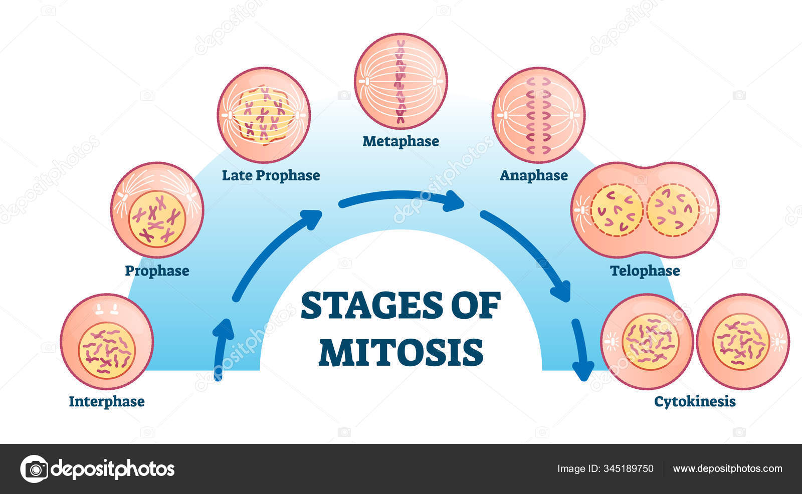
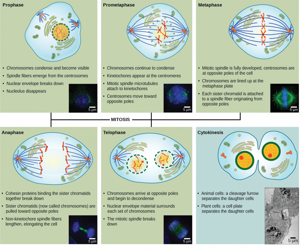
These are the stages of Mitosis, including pictures, illustrations, and explanations of each stage, excluding cytokinesis. Figure: Stages of mitosis. Image Source: Wikipedia (Ali Zifan). Mitosis is divided into the following phases based on the completion of one set of activities and the onset of the other. 1. Interphase. Interphase is a part of the cell cycle where the cell copies its DNA as preparation for the M phase (mitotic phase). Figure 1 - Microscope image of cells in various stages of mitosis. Stages of Mitosis Prophase . Each chromosome is made of two genetically identical chromatids, joined by a centromere. During DNA replication, genetic material is loosely packed as chromatin. However, during mitosis DNA needs to be more tightly packed to allow for easier

The 4 Mitosis Phases: Prophase, Metaphase, Anaphase, Telophase Biology Diagrams
#2: "Mitosis: Splitting Up Is Hard To Do" by Crash Course If you're a bit exhausted from reading dense material and need someone else to put the stages of mitosis into more accessible terms, head over to YouTube and watch Crash Course's 10 minute video on mitosis, called "Mitosis: Splitting Up Is Hard to Do.". The nice thing about this video is that, while being a bit more thorough Stages of mitosis. Karyokinesis (or mitosis) is divided into five stages—prophase, prometaphase, metaphase, anaphase, and telophase. The pictures at the bottom were taken by fluorescence microscopy (hence, the black background) of cells artificially stained by fluorescent dyes: blue fluorescence indicates DNA (chromosomes) and green Before the cell enters the mitosis phase, it first undergoes a synthesis or S phase where each chromosome is duplicated and consists of two sister chromatids joined together by a specific DNA sequence known as a centromere. Centromeres are crucial to segregation of the daughter chromatids during mitosis. No images, graphics, software