UNIT 9 JOINTS elbow joint diagram Diagram Biology Diagrams The proximal radioulnar joint is a separate articulation within the elbow joint capsule. Protrusions and Bony Prominence Olecranon: The pointy bony structure at the posterior side of the elbow; it is the protrusion at the upper end of the ulna

The elbow joint, although non-weight bearing, may be the most complex joint in the human body. The elbow is a synovial hinge joint made up of articulations of mainly the distal humerus and the proximal ulna. However, articulations exist between the proximal radius and the humerus as well as the proximal radius and ulna. The three articulations are referred to as the ulnohumeral, radiohumeral

Elbow Joint Anatomy: A Comprehensive Guide to Osseous Structure Biology Diagrams
first motor branch to FCU is found distal to the elbow joint. Blood Supply of Elbow. Brachial artery. is located medially in the upper arm. Diagnosis, Treatment and Rehabilitation. CHAPTER: ELBOW ANATOMY Francesc Malagelada, Miki Dalmau-Pastor, Jordi Vega, Pau Golano. Shoulder & Elbow - Elbow Anatomy & Biomechanics The elbow represents a complex hinge joint that combines stability with mobility to enable essential upper extremity functions. This intricate articulation between the humerus, radius, and ulna facilitates both flexion-extension movements and forearm rotation, making it crucial for activities of daily living and specialized tasks. Like all synovial joints, the elbow joint has a capsule enclosing the joint. This is strong and fibrous, strengthening the joint. The joint capsule is thickened medially and laterally to form collateral ligaments, which stabilise the flexing and extending motion of the arm. A bursa is a sac-like structure containing a small amount of synovial
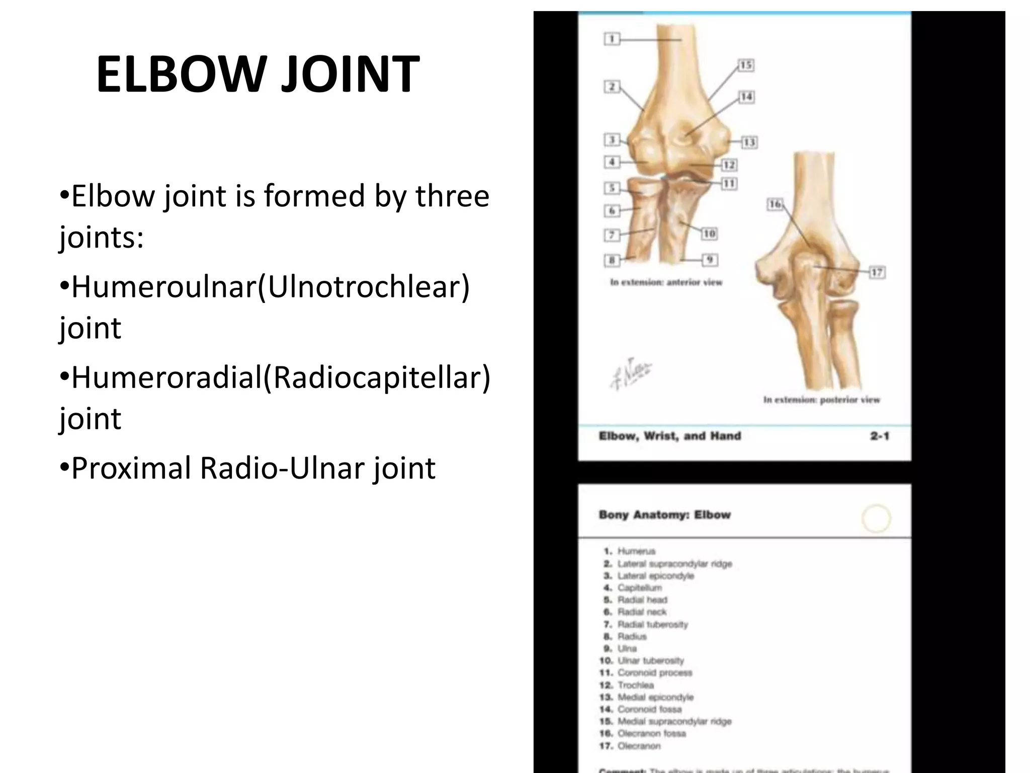
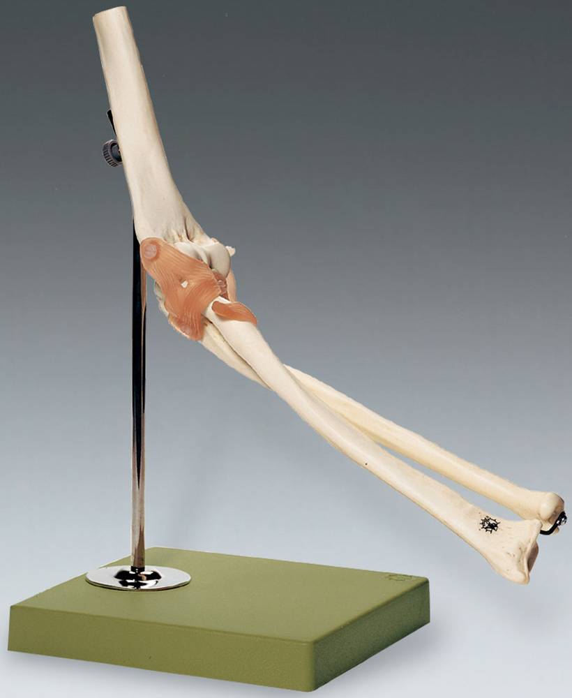
The elbow joint is a synovial joint that connects the arm and the forearm, providing 150 ْ of extension-flexion movement. It consists of three joints; the humeroulnar joint, the humeroradial joint, and the proximal radioulnar joint, all within one articular capsule! The elbow joint is supported by three ligaments: The annular ligament
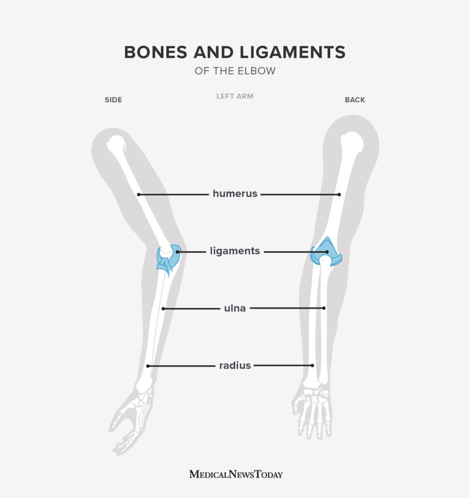
Elbow Bones: Names, Basic Anatomy, & Diagrams Biology Diagrams
The elbow joint is a vital structure for upper limb function, making it prone to injuries and disorders due to its role in movement, weight-bearing, and stability. Fractures. Common fractures involve the distal humerus, radial head, or olecranon process of the ulna, often caused by falls or direct trauma.